10 FREE IMAGES of ShutterstockTry Shutterstock and Get 10 Free Images

conidia SVG, Vector And Clipart Images

() conidia SVG Vectors and Transparent background conidia ClipArts. All Download the royalty free graphic in the form of SVG, PNG, EPS, AI or PSD.

Description:

Usage ideas:

Structure of Penicillium. Mycelium with conidiophore and conidium isolated on white background clipart svg vector

Structure of Penicillium. Mycelium with conidiophore and conidium isolated on white background

Reproductive Structures of Penicillium. Life cycle clipart svg vector

Reproductive Structures of Penicillium. Life cycle

Structure of Penicillium. Mycelium with conidiophore and conidium isolated on white background clipart svg vector

Structure of Penicillium. Mycelium with conidiophore and conidium isolated on white background

Penicillium Slide ( blue mold, mycelium and conidiophores). Mold Microscopy clipart svg vector

Penicillium Slide ( blue mold, mycelium and conidiophores). Mold Microscopy

Structure of Penicillium. Mycelium with conidiophore and conidium isolated on white background clipart svg vector
Structure of Penicillium. Mycelium with conidiophore and conidium isolated on white background

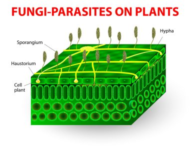
The parasitic fungi absorb their food material from the living tissues of the hosts on which they parasitize. Pathogens. education Vector diagram clipart svg vector

The parasitic fungi absorb their food material from the living tissues of the hosts on which they parasitize. Pathogens. education Vector diagram

Penicillium anatomy. Structure of a Microscopic fungi that use in food and drug production. Part of a Fungus. Close-up of a Metula, Sterigma, Conidia, Hypha. vector illustration isolated on white background. clipart svg vector

Penicillium anatomy. Structure of a Microscopic fungi that use in food and drug production. Part of a Fungus. Close-up of a Metula, Sterigma, Conidia, Hypha. vector illustration isolated on white background.

An image shows mycelium. This image shows an enlarged view of the mycelium. a: Ascocarp or Perithecium, c: erect Hyphae forming spores or Conidia, h: Haustoria penetrating epidermis of leaf, vintage line drawing or engraving illustration. clipart svg vector

An image shows mycelium. This image shows an enlarged view of the mycelium. a: Ascocarp or Perithecium, c: erect Hyphae forming spores or Conidia, h: Haustoria penetrating epidermis of leaf, vintage line drawing or engraving illustration.
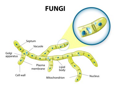
Typical fungi cell. Fungal Hyphae. Structure fungi. clipart svg vector

Typical fungi cell. Fungal Hyphae. Structure fungi.

Structure of Penicillium. Mycelium with conidiophore and conidium isolated on white background clipart svg vector

Structure of Penicillium. Mycelium with conidiophore and conidium isolated on white background

Structure of Penicillium, Opportunistic fungi that cause mucormycosis involving skin under the optical microscope, isolated on white background. clipart svg vector

Structure of Penicillium, Opportunistic fungi that cause mucormycosis involving skin under the optical microscope, isolated on white background.

Mold life cycle. The structure of mold. clipart svg vector

Mold life cycle. The structure of mold.

Penicillium Mold Microscopic Illustration. Detailed Penicillium Fungus Spore Structure. Scientific Visualization of Mold Growth clipart svg vector

Penicillium Mold Microscopic Illustration. Detailed Penicillium Fungus Spore Structure. Scientific Visualization of Mold Growth