Reproductive Structures of Penicillium. Life cycle

Structure of Penicillium. Mycelium with conidiophore and conidium isolated on white background

Penicillium Slide ( blue mold, mycelium and conidiophores). Mold Microscopy

Structure of Penicillium. Mycelium with conidiophore and conidium isolated on white background

Cordyceps Engleriana, Perithecium and Conidium on the spider. Publication of the book "Meyers Konversations-Lexikon", Volume 7, Leipzig, Germany, 1910

Penicillium anatomy. Structure of a Microscopic fungi that use in food and drug production. Part of a Fungus. Close-up of a Metula, Sterigma, Conidia, Hypha. vector illustration isolated on white background.

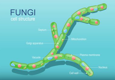
Anatomy of typical fungi cell. Fungal Hyphae and Cell Structure. Vector diagram

Structure of Penicillium. Mycelium with conidiophore and conidium isolated on white background

Structure of Penicillium. Mycelium with conidiophore and conidium isolated on white background

Structure of Penicillium, Opportunistic fungi that cause mucormycosis involving skin under the optical microscope, isolated on white background.

Mold life cycle. The structure of mold.